


The reasons people start looking into lateral canthoplasty revision are usually clear. After lateral canthoplasty, the outer corner may fail to hold its tapered shape and spread into what is called a webbing deformity, or the red inner lining may become visible as scleral show. Both damage the balance of the eye, functionally and aesthetically. When the deformity is severe on one side only rather than both, the asymmetry stands out and the daily discomfort patients feel grows considerably.
In that situation, revising only the affected side to match the intact side is often the more anatomically sound approach, rather than operating on both eyes for the sake of symmetry. This article looks at the medical reasoning behind a case in which severe lateral canthoplasty side effects on one eye were corrected with a single-side revision, followed over two months.
What a single-side revision actually looks like
A case from our official Naver blog. The full photo series is there.
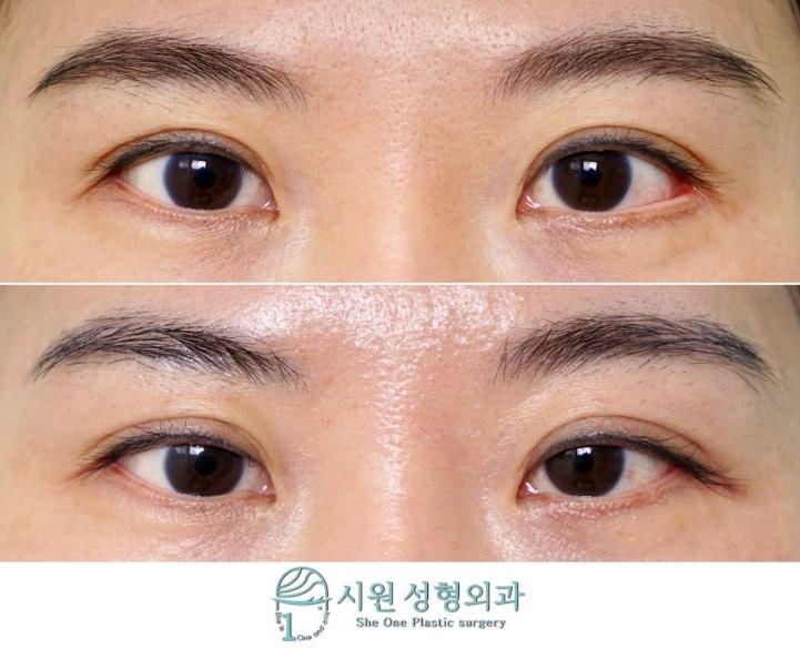
The before and after photos in this post are published with the patient’s consent.
If you assumed both eyes had to be done — two-month course, 35 photos ›
Why a single-side reconstruction can be the right choice
Eye surgery is often performed on both sides at once so that the two can be matched. But when a previous operation has damaged the gray line of one outer corner so badly that the lining has everted, the approach has to change. Here the eye whose structure is still intact becomes the reference point, and only the damaged side is adjusted. That places less strain on the outer corner and is the most objective way to reduce the asymmetry.
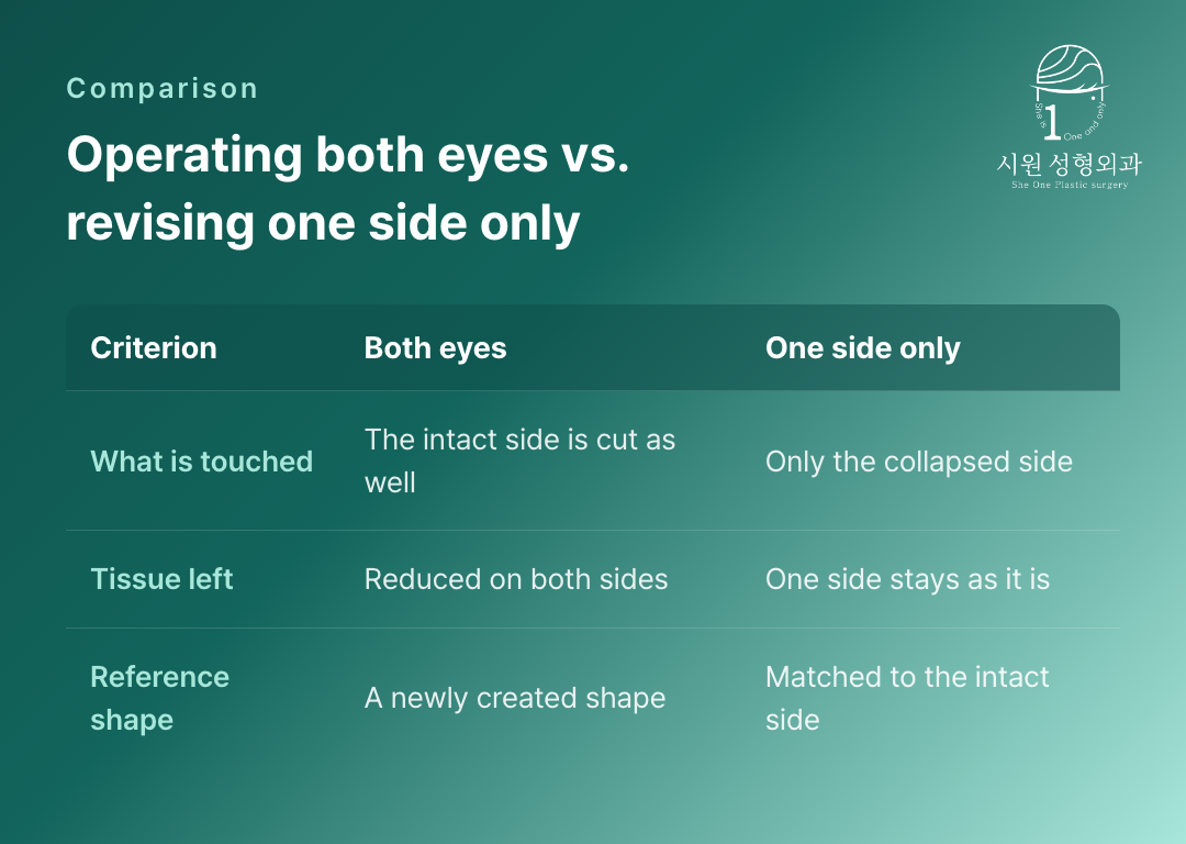
Why a single-side revision was chosen over a combined approach
This patient’s priority was not to preserve horizontal eye length at all costs. It was to close the exposed red lining, relieve the functional discomfort, and bring the height difference between the two outer corners back toward its original form. The surgery therefore concentrated on tapering the outer corner again and re-anchoring the lateral canthal ligament firmly to the orbital bone, stabilising the collapsed structure.
How the tissue settles at day 8 and at two months
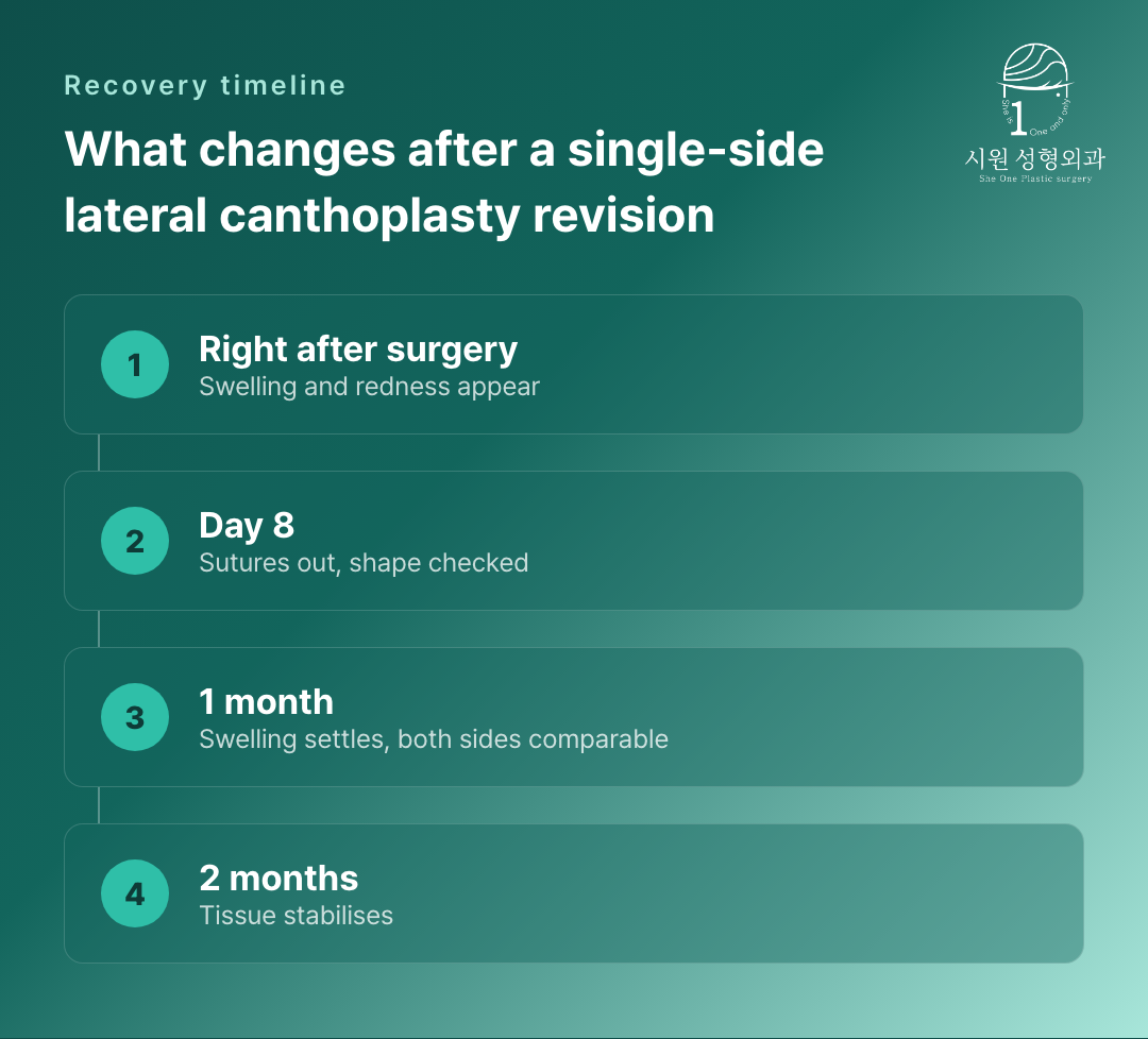
At day 8, when the sutures come out, the tension of the outer corner being anchored to bone produces temporary tightness, stiffness and slight swelling at the surgical site. This is an unavoidable early response to fixing an everted lining closed. By around two months the internal scar tissue softens and the structure settles into place. In this patient, the lining at the left outer corner that had been red and open had come to sit against the globe, and the asymmetry had eased so that the slope resembled the intact right side.
Assessing the structural limits honestly
When correcting eyes affected by side effects, the same operation cannot be applied uniformly to every case. The tension of the muscles around the eye and the condition of the damaged ligament have to be assessed objectively so that a reasonable plan can be built, one that does not strain the underlying framework. If you are troubled by a markedly asymmetric outer-corner deformity, a detailed examination that identifies the cause precisely is the sensible starting point for a realistic structural improvement.
The before and after photos featured in this post are shared with the patient’s explicit consent in accordance with the Medical Service Act. Individual recovery times and results vary, and side effects can occur. Please consult our medical team before making any surgical decision.
Official WebsiteProcedures and medical staff›
KakaoTalk ConsultationSend us photos and ask›
LINE ConsultationEasier if you are outside Korea›
DirectionsApgujeong Stn. · JJ Building 2F›
2F, JJ Building, 19 Apgujeong-ro 28-gil, Gangnam-gu, Seoul, Korea
Mon–Fri 10:00 – 19:00 · Sat 10:00 – 16:00
Closed Sun & public holidays · 02-512-1889
