

The two directions each procedure lengthens
Many people go in to ask about a lateral canthoplasty — an opening at the outer corner — and come away having also been offered a lower-corner opening. If you are unsure whether both are really needed, you are not alone.
That recommendation is not simply a matter of adding one more operation. It starts from an anatomical fact: the structure that forms the outer corner is bound into one piece with the lower eyelid. Moving the lateral canthus outward, or bringing it down, means working on the lateral canthal tendon that holds the upper and lower tarsal plates together, so it is difficult to plan in complete isolation from where the lower lid sits. Adjusting only the horizontal length limits what is actually noticed from the front; adjusting only the vertical height does not change how long the eye reads. The two axes have to be considered together.
What follows sets out why the two are carried out together, and equally the cases in which they need not be.
What does each of the two procedures lengthen?
The names are similar, but the axis each one works on is different.
An opening at the outer corner is a lateral canthoplasty: the outer corner is incised and the lateral canthus extended outward, lengthening the eye horizontally.
An opening at the lower corner corresponds to lower eyelid lowering: roughly the outer third of the lower lid is brought down, widening the eye vertically. The lower part of the iris that had been covered becomes visible, and the eye looks larger.

The lateral canthal tendon and the structures of the lower lid
Why are the two offered together?
The reason is not that two procedures simply make the eye bigger. The outer corner and the lower lid share one supporting structure, so adjusting one alone leaves the border of the other out of line. That is the technical ground for combining them.
What does the change look like in numbers?
A cohort that adjusted the horizontal and vertical axes in a single combined canthoplasty (82 East Asian women, 164 eyes, mean age 30.06) reported the following measured values six months after surgery.
- Pupil centre to outer corner (horizontal) — 12.68 mm before, 14.63 mm after
- Canthal tilt — 7.95 degrees before, 5.77 degrees after
- Pupil centre to lower lid margin (vertical) — 5.70 mm before, 6.75 mm after
- Pupil centre to lower lid margin at the lateral limbus — 4.95 mm before, 5.85 mm after

Measured change at six months after a combined procedure
Are there eyes for which the outer corner alone is enough?
- It depends on the technique. With an incisional lateral canthoplasty the upper and lower lid lines are separated, so bringing the outer corner downward is described as requiring a lower-corner opening alongside. A V-shaped approach, which moves the corner point itself, can bring the corner down without one.
- The cause may not lie at the corners at all. Where a heavy look comes from a weakness in the lifting force — that is, ptosis — it has been pointed out that ptosis correction should be considered before any opening. An opening is not a compulsory part of double-eyelid surgery.
- Some eyes do not lengthen well. Where the canthal tilt is 10 degrees or more, or the lateral fornix depth is 2 mm or less, the lengthening effect of an outer-corner opening has been reported to be relatively small. With a fornix depth of 2 mm or less, concentrating on lowering the corner and deepening the fornix is recommended as safer than forcing the length.
In other words, “as large as possible” should not be the goal. An accurate diagnosis of whether the two procedures are genuinely called for has to come first.
What has to be watched when they are done together?
Because the scale of the change is larger, a problem shows more plainly — a point made repeatedly in Korean specialist writing. Limits that must not be crossed are given alongside: the outer-corner opening cannot pass the orbital rim, and the lower-corner opening cannot pass the lower edge of the iris. Past those lines, red tissue and sclera show.
Where the structure loses its support, the following changes are reported.
- Sclera showing below the iris — white appears under the iris, which can give an older rather than a larger-eyed impression.
- Lagophthalmos — the eye no longer closes fully, bringing dryness and other discomfort.
- Ectropion — arising from shortening of the anterior lamella, the lower lid falls away from the globe.
- A blunted or rounded outer corner, a squared-off deformity, mucosal exposure — from excessive tightening or a suture placed wrongly.
What deserves attention is that these do not appear separately from one another.
That said, there is no ground for asserting that doing both at once multiplies the complications. No study directly comparing complication rates between simultaneous and staged surgery was found. It is closer to the truth to understand it as a structure in which the result is simply more visible, because the change is larger — not one in which the number of complications rises.
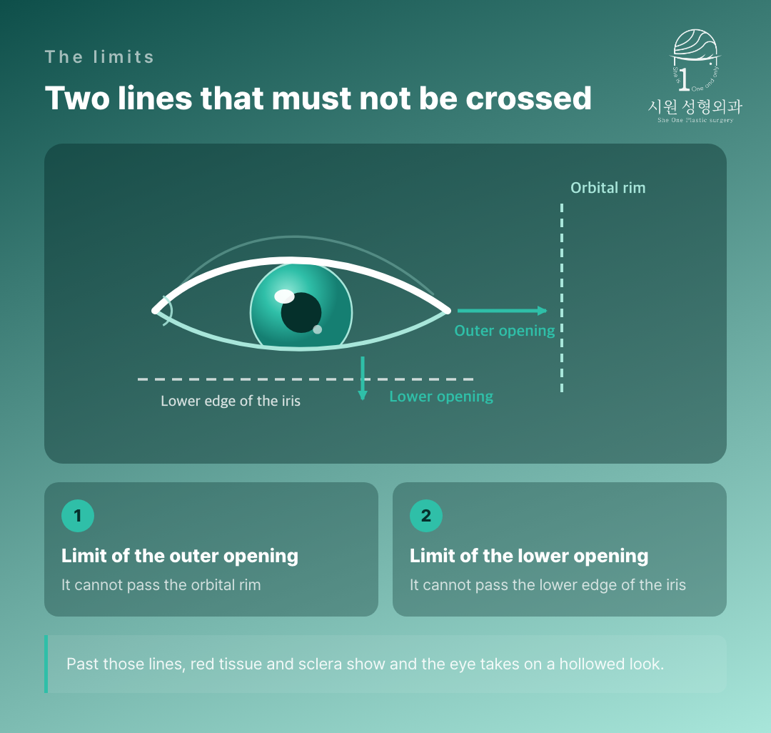
The two limits, and what happens past them
What is checked in consultation?
The items below gather conditions recorded in the literature. They are not a self-diagnosis tool; they are what is checked together with measurement at consultation.
- Whether the lateral fornix depth is 2 mm or less — a condition in which horizontal lengthening does not take well.
- Whether the canthal tilt is 10 degrees or more — the lengthening effect is reported to be relatively small.
- Whether the palpebral fissure is 25 mm or less, or the upward tilt 15 degrees or more — conditions under which ectropion, scarring, webbing and conjunctival exposure were frequent with traditional techniques.
- Whether a weakness in the lifting force — ptosis — is present alongside; ptosis correction may take priority over any opening.
- Whether there is a negative canthal tilt — described as carrying a significantly higher risk of lid malposition after surgery.
- Whether the globe is prominent and there is laxity of the tarsus and tendon — a condition associated with sclera showing.
On the ideal height of the lateral canthus, sources divide. Some describe it as about 2 mm above the medial canthus; others propose about 4.1 degrees, or 1.2 mm above, and mention an upward tilt of up to 3 mm. The basis of measurement itself differs, so a single figure cannot be settled on. It is an area to be judged within the balance of the individual face rather than by aiming at a number.

What is checked, together with measurement, in consultation
Questions we are often asked
Why am I told to have both done together?
Because the two lengthen in different directions. An outer-corner opening takes care mainly of the horizontal width and a lower-corner opening of the vertical height, and given the anatomy of the eye the two work together when carried out at the same time.
Can I have the lower-corner opening on its own?
If the aim is vertical widening or a softer impression, yes — but it will not lengthen the eye horizontally. Where the change you want is limited to the vertical, it is a reasonable option on its own, and there is no need to widen the scope unnecessarily.
If I have both, are the complications doubled?
No study calculating a doubled rate was found. What is pointed out is that a simultaneous procedure makes a problem more visible because the change is larger, and that in cases that went wrong, corner deformity, lower-lid retraction, sclera showing, ectropion and lagophthalmos tended to appear together. We do not present only the results from when things go well; we explain how it looks when it fails, and set the scope after that.
How does recovery go?
Sutures usually come out at seven to nine days. Bruising settles in seven to ten days and swelling within about a month, while redness along the incision can remain for up to three months. For three weeks, rubbing the eye or any impact to it should be avoided. Recovery speed varies between individuals, so the course has to be followed case by case.
In closing
We understand how long patients weigh the wish for large, open eyes against the worry that the change might go too far and look unnatural.
We do not recommend combining procedures beyond what is needed. Through a one-to-one consultation we assess the length of the palpebral fissure, the canthal tilt and the supporting condition of the lower lid objectively, and propose only the scope that can be carried out safely and without strain — opening only as much as is needed, and keeping the structures that have to be kept.
Individual recovery times and results vary, and side effects can occur. Please consult our medical team before making any surgical decision.
WhatsApp ConsultationInternational patients welcome›
Official WebsiteProcedures and medical staff›
KakaoTalk ConsultationSend us photos and ask›
LINE ConsultationEasier if you are outside Korea›
DirectionsApgujeong Stn. · JJ Building 2F›
Consultations are held in English with the surgeon and our staff directly, not through an interpreter. Written enquiries in English are welcome.
2F, JJ Building, 19 Apgujeong-ro 28-gil, Gangnam-gu, Seoul, Korea
Mon–Fri 10:00 – 19:00 · Sat 10:00 – 16:00 · (KST, UTC+9)
Closed Sun & public holidays · +82-2-512-1889
