
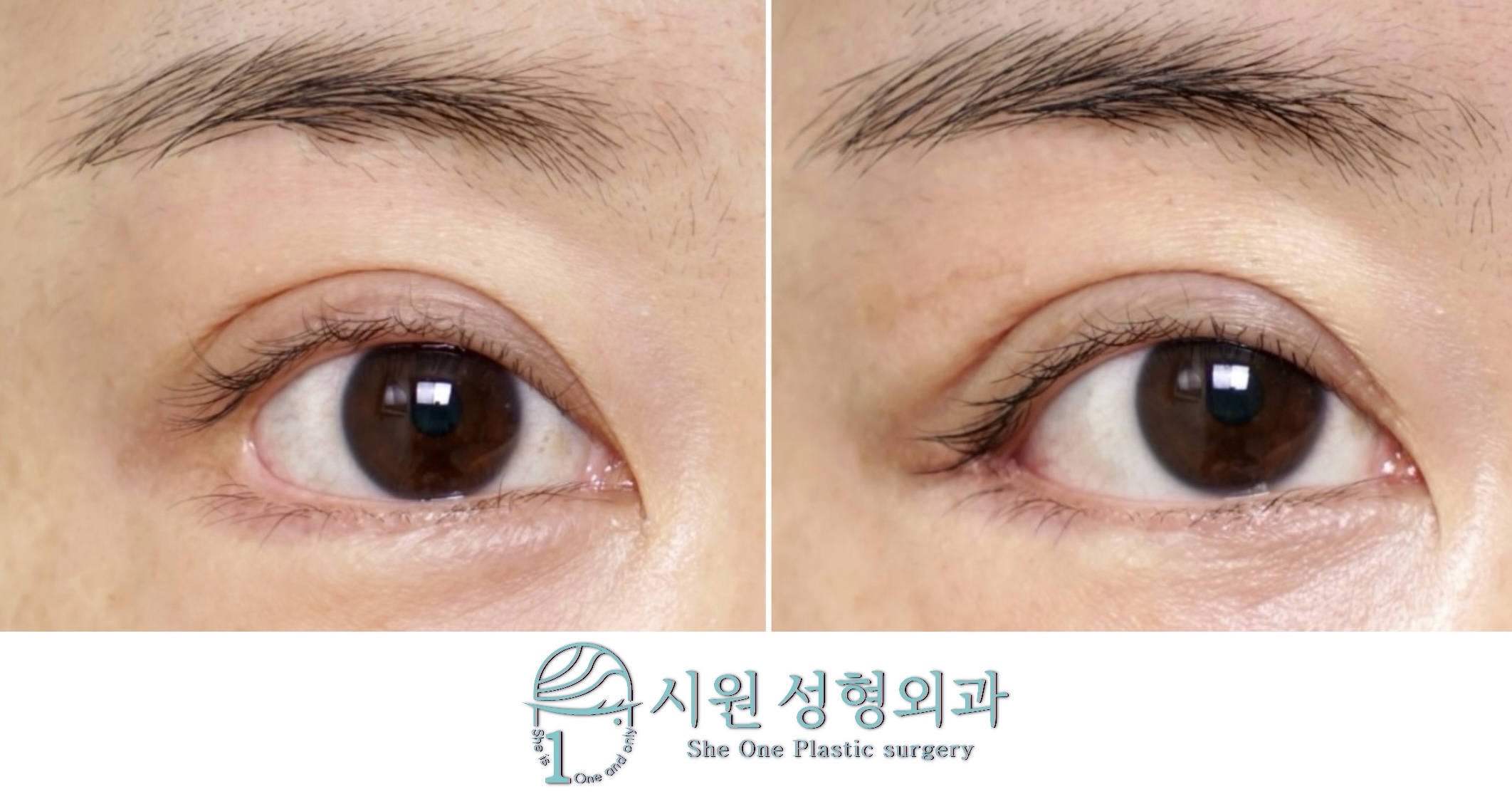
When eye surgery chosen in the hope of a larger, clearer eye instead leaves the outer corner gaping or the whole look unnatural, the distress is hard to put into words. Reconstruction after a lateral canthoplasty is the operation that reverses that state.
More people are presenting with the outer border of the eye having given way after an indiscriminate corner opening — mucosa showing red, or the outer corner dropping so that the face reads as downcast. This kind of collapse does not heal of itself with time, so a precise reconstruction that returns the anatomy to where it was is needed.
What follows traces, through one patient’s course at three weeks after reconstruction of the outer corner, how damaged lateral canthal tissue is restored structurally.
The course at three weeks, in photographs
A case from our official Naver blog. Before, day 8 and month 2 are compared there.
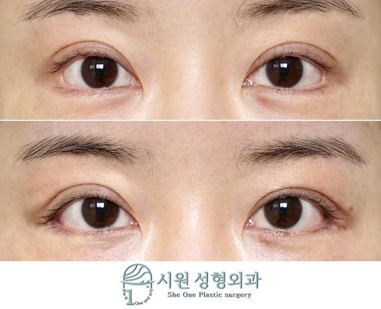
The before and after photos in this post are published with the patient’s consent.
An outer corner gaping into a squared shape and turned downcast — the course at three weeks ›
1. What deforms an outer corner, and why reconstruction is needed
The outer corner of the eye — the lateral canthus — is a point where bone, ligament and fine muscle interlock. If the supporting structure beneath the skin is not secured firmly at the first operation and only the surface skin is pulled and excised, the site cannot withstand the tension of eye movement and gradually gives way.
The result is an end that no longer comes to a sharp point but looks blunt and hollowed, with the conjunctiva — which should sit against the globe — exposed, bringing functional discomfort such as dry eye. And as the lateral canthal tendon loses the strength with which it held the lower lid taut, the whole outer corner sinks, forming an eye that reads as tired and sad without meaning to.
2. Restoring the anatomical order
To improve a state in which function and appearance have been damaged together, we avoid the one-dimensional approach of simply suturing the gap closed.
- Returning the canthal tendon to its place — after separating the internal adhesion knotted with scar tissue delicately, the tendon that has lost its hold and slipped is located and re-fixed firmly at its normal position on the orbital bone.
- Layer-by-layer restoration — not the surface alone but the mucosa and the muscle layer beneath are separated and sutured in several layers, each according to its own order. This draws the collapsed corner back into a sharp form and lifts the dropped angle naturally.
3. Three weeks on: early stability
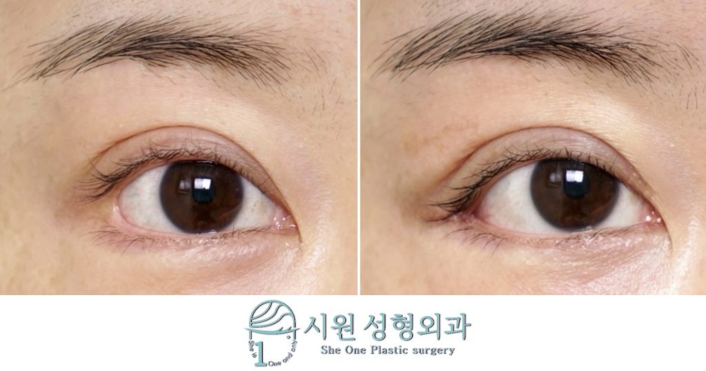
The course at three weeks after reconstruction of the outer corner
This is the appearance at three weeks, with an outer corner that had been blunt, gaping and dropped now tidied into a sharp form.
In revision surgery, the tissue usually needs several months to soften and for the shape to settle fully. Even so, at only three weeks after surgery, the corrected line of the outer corner in this case is holding structurally without noticeable movement.
The mucosa that had been exposed red is comfortably covered again, and the axis of the outer corner, which had fallen and read as sad, has returned to within its normal range for a more harmonious and softer impression. This is the kind of stable recovery that can be expected when unnecessary tissue damage during surgery is reduced and the technique is fitted to the patient’s own anatomy.
Back to the ordinary
We know better than anyone the loss and the fear a patient carries in lying down on the operating table once again after a painful experience of failure.
Rather than overcorrecting out of ambition, we propose only the safest and most natural solution the patient’s present tissue can carry. We give our full care to every step, so that a long-standing hurt can heal and this choice becomes a true full stop to a tiring journey.
The before and after photos featured in this post are shared with the patient’s explicit consent in accordance with the Medical Service Act. Individual recovery times and results vary, and side effects can occur. Please consult our medical team before making any surgical decision.
WhatsApp ConsultationInternational patients welcome›
Official WebsiteProcedures and medical staff›
KakaoTalk ConsultationSend us photos and ask›
LINE ConsultationEasier if you are outside Korea›
DirectionsApgujeong Stn. · JJ Building 2F›
Consultations are held in English with the surgeon and our staff directly, not through an interpreter. Written enquiries in English are welcome.
2F, JJ Building, 19 Apgujeong-ro 28-gil, Gangnam-gu, Seoul, Korea
Mon–Fri 10:00 – 19:00 · Sat 10:00 – 16:00 · (KST, UTC+9)
Closed Sun & public holidays · +82-2-512-1889
