
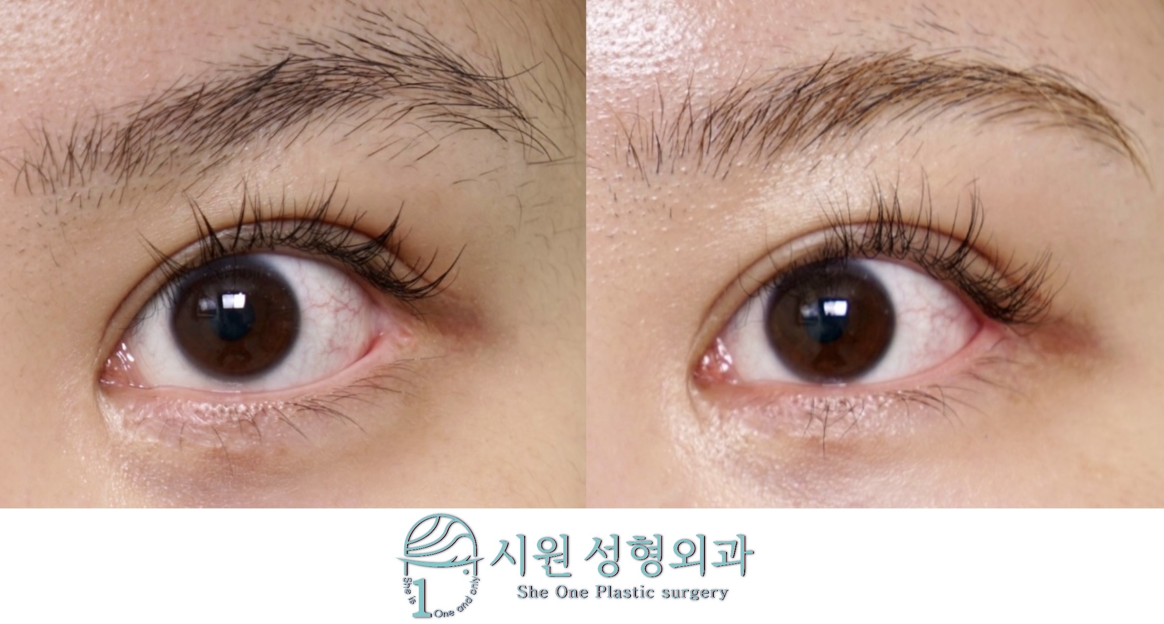
A lateral canthoplasty side effect can appear when surgery expected to create large, defined eyes leaves unintended results, and the psychological distress that follows is considerable.
When the structure supporting the end of the eye is lost due to excessive surgical manipulation, the outer corner, which should originally be sharp, deforms bluntly, and the internal red mucosa appears flipped outward. This phenomenon is not merely an aesthetic concern but is accompanied by functional deterioration such as dryness or a stinging sensation on the ocular surface. Since damaged tissues cannot heal simply with time, a precise reconstruction process that returns the anatomical structure to its proper position must accompany it.
Today, Sheone Plastic Surgery introduces a 2-month progress case that erased the traces of a previous surgical failure and transformed into a comfortable impression.
Pre-operative Symptoms and Diagnosis Details
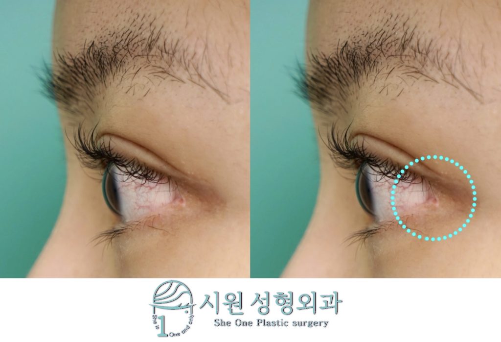
Precise analysis of outer corner deformation for lateral canthoplasty side effect revision
Condition at the time of visit : Due to the previous surgery, the supporting base around the lateral canthus weakened, causing the end of the eye to gape in a round and blunt shape. As a result, red mucosal exposure is clearly observed, and the scar tissue near the outer corner stands out, making the overall eye shape look awkward and sharp.
Patient’s concerns and needs : The patient wanted to hide the scar by pushing the outwardly flipped mucosa inward, and to return the roundly deformed outer corner shape to the sharp and smooth V-line from before the surgery.
Sheone Plastic Surgery’s solution : Lateral canthoplasty revision + Revision canthoplasty + Outer corner styling (height adjustment)
Correcting a Lateral Canthoplasty Side Effect: Ligament Re-anchoring and Recovery Stages
A method that simply forces the skin at the end of the eye to be pulled and sutured cannot overcome the physical tension applied when opening and closing the eyes. This highly likely leads to wound gaping or secondary deformation. At our clinic, we microscopically separate the skin, muscle, and scar tissue layer by layer, then locate the severed lateral canthus ligament and firmly anchor it to the periosteum to perform a structural reconstruction.
We cannot newly grow the hair follicles in the area where eyelashes were lost due to the previous failure. However, since the loosened tissue is pulled tightly at a normal angle, the empty space without eyelashes is physically reduced, correcting it so that it is visually unnoticeable.
Because the severed lateral canthus ligament is firmly bound to the bone, you may feel a somewhat pulling sensation when tightly closing your eyes or making facial expressions during the early stages of surgery. This is a highly natural phenomenon that occurs in the process of the tissue adhering to its proper place, and it gradually decreases as the scar softens over about 3 to 4 months.
Careful attention is required during the period when chemosis and swelling appearing immediately after surgery recover. Stitches are generally removed around 7 to 10 days, and you can do a light water wash from the next day. To reduce irritation to the surgical site, we recommend proceeding cautiously with eye makeup (such as eyeshadow) starting 2 to 3 weeks after the stitch removal point.
2-Month Post-operative Progress and Change Verification
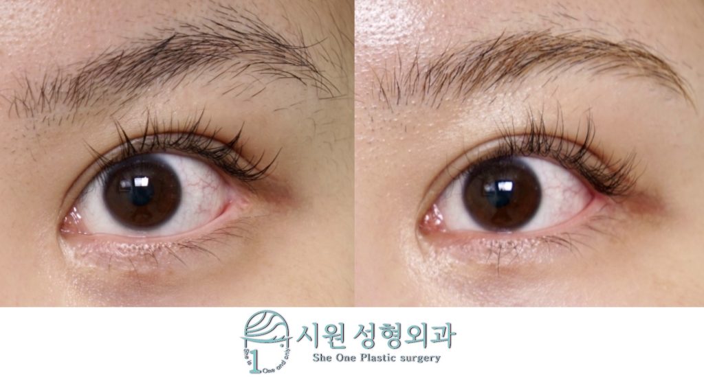
2-month progress of blunt outer corner deformation and mucosal exposure correction
As 2 months pass after the surgery, a significant portion of the swelling has subsided, and the corrected tissues have entered a stabilization phase.
Improvement of outer corner gaping and deformation : The end of the eye, which had lost its taut supporting power and gaped bluntly, was corrected into its original sharp and sophisticated V-shape through precise lateral canthus re-anchoring.
Improvement of mucosal exposure and scars : The mucosal exposure area, which was flipped outward giving visual fatigue, solidly entered its proper anatomical position. Along with the refined scars, you can confirm that the somewhat sensitive impression before the surgery has improved into a much softer and natural eye shape.
Customized Care Correcting the Anatomical Structure
Sheone Plastic Surgery deeply empathizes with the emotional distress you must have experienced, finding it difficult to even make eye contact with others due to a damaged outer corner.
Eye revision surgery should not stop at merely suturing the visible epidermis; it is a highly difficult surgical technique that must simultaneously draw out functional and aesthetic stability by restoring the damaged internal structure. Sheone Plastic Surgery strictly avoids reckless overtreatment and proposes only safe surgical plans that correspond to each individual’s tissue condition through a 1 : 1 precise analysis. We promise honest care at all times so that those who step in with exhausted hearts can regain a comfortable and clear gaze.
* Before and after photos of this patient are available on the She One Plastic Surgery official blog. *
See the actual 2-month progress
A case from our official Naver blog. See the full photo series.
The before and after photos featured in this post are shared with the patient’s explicit consent in accordance with the Medical Service Act. Please note that individual recovery times and surgical results may vary, and potential side effects can occur. We strongly advise a comprehensive consultation with our medical team before making any surgical decisions.
Official WebsiteProcedures and medical staff›
KakaoTalk ConsultationSend us photos and ask›
LINE ConsultationEasier if you are outside Korea›
DirectionsApgujeong Stn. · JJ Building 2F›
2F, JJ Building, 19 Apgujeong-ro 28-gil, Gangnam-gu, Seoul, Korea
Mon–Fri 10:00 – 19:00 · Sat 10:00 – 16:00
Closed Sun & public holidays · 02-512-1889
