
What makes an epicanthoplasty look done?
The worry we hear most from people who want a natural epicanthoplasty is that it will look operated on. In consultation the question is usually how much to open — but whether it shows is decided by the direction of the opening and by the conditions of that particular eye, not by the amount.
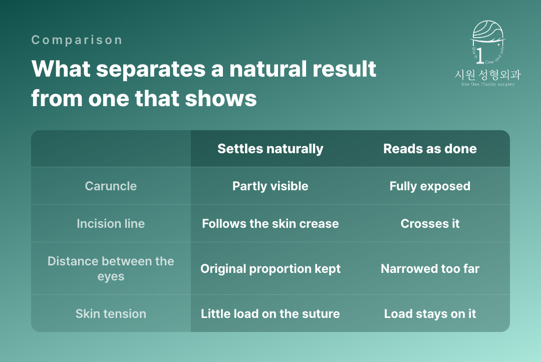
Opening the inner corner reveals three things at once: the red caruncle, the incision line, and a changed distance between the eyes. If any one of the three falls outside the proportions of the face, the eye looks larger but the work shows.
What follows looks at what separates a natural epicanthoplasty from one that reads as done, and at what is measured before surgery.
How does the thickness of the fold change the result?
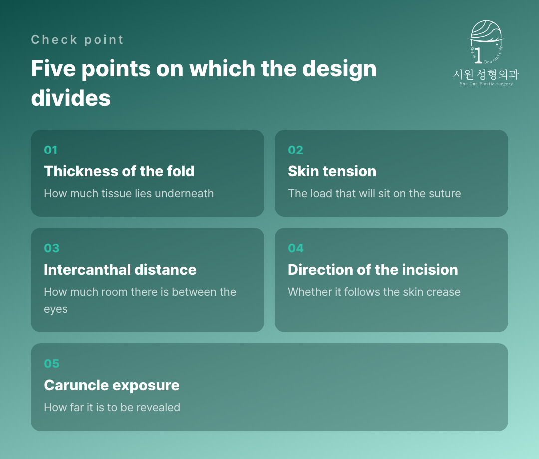
The epicanthal fold differs in thickness and in shape from person to person. According to a plastic surgery report analysing the shape and size of the fibrous band at the medial canthus, differences in that band govern the degree and the type of fold. The size of the fold as seen from outside does not tell you about the structure beneath.
An eye with a thick, firm fold has more tissue to release underneath, even for the same length of incision. A thin fold, by contrast, exposes the caruncle markedly with only a small opening, so the width has to be kept back instead. Setting the length without looking at the thickness sends the result wrong in one direction or the other.
Thickness is hard to judge by eye alone. We check how far the fold follows when it is lifted gently with a fingertip and how it resists from within, and translate that into the depth to be released rather than into a length of incision.
Why does skin tension govern the scar?
Where load keeps acting on a sutured site, the scar settles wide and thick. The inner corner is pulled every time the eye opens and closes, so that force acts throughout recovery.
So when the incision is designed, the direction in which tension is relieved is considered alongside. A plastic surgery report on incisions that release tension in the skin at the inner corner exists as a subject in its own right — where the force is led is one axis of epicanthoplasty design. Reports on reducing scarring likewise focus on tension and the extent of dissection rather than on how much is incised.
Can the direction of the incision make the scar show?
Skin has its own lines along which it folds. An incision that follows those lines hides within them; one that crosses them shows as a line, at the same length. At a site that moves as much as the inner corner, that difference is especially marked.
The area dissected matters too. A craniofacial surgery report covering 421 epicanthoplasties notes that dissecting more widely than necessary can leave a hypertrophic scar or a web-shaped deformity. Where a depressed scar has already settled across a wide area, restoring it in stages can be safer than covering it in one go.
Care during recovery tells the same story. Avoiding sunlight and not disturbing the wound can help slow a scar from darkening, but no amount of care removes the force already acting on the suture line. The direction of that force is set in the surgical design.
Why is the intercanthal distance measured beforehand?
The intercanthal distance is the gap between the inner ends of the two eyes. An epicanthoplasty acts to narrow it, so a face with a generous gap has room to spare, while in one that is already narrow even a small opening makes the eyes look crowded.
That is why the judgement has to be made within the proportions of the whole face. A width decided by looking at the eyes alone can satisfy up close in the mirror and read as odd in a photograph of the face. Keeping the width sparing at a first operation is what leaves the later options open.
What do results that show have in common?
Among people who come to us for correction there is a pattern. The caruncle is exposed to a degree that makes the eye look severe; the incision has left a white line or a depression; or the two eyes look crowded together. All three arise from direction and extent, not from the amount opened.
Undoing is a harder proposition than opening in the first place, because the skin to spare has shrunk and scar tissue has accumulated layer by layer. Why a revision falls short usually begins here, which is why leaving a margin in the first plan is the safer course.
- The caruncle is fully exposed and the red part is visible from the front.
- The incision has left a white line or a depressed mark.
- The distance between the eyes has narrowed and the face reads as crowded.
- A hook-shaped fold has formed below the inner corner.
- The two inner corners have settled into different shapes.
What to establish in consultation
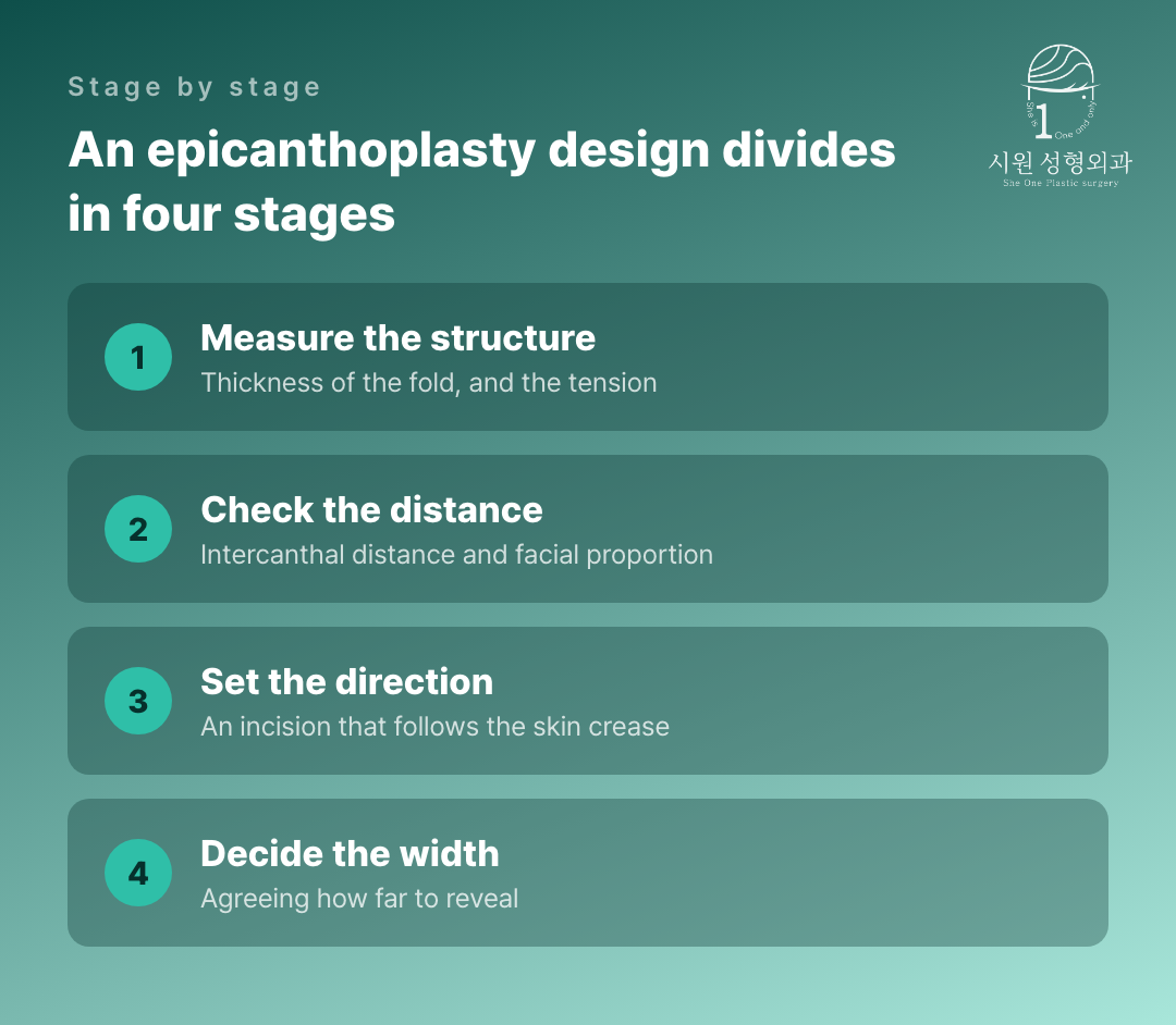
Rather than how many millimetres will be opened, it helps to ask whether the thickness of your fold and your intercanthal distance were actually measured, and what width those values pointed to.
A natural epicanthoplasty is not settled by the name of a particular technique. The same method gives different results on a thick fold and on a thin one. If an explanation stays with the name rather than the measurement, it is worth asking once more.
An inner corner opened too far is among the harder things to undo, which is why the question we spend longest on is not how much can be opened but how much should be.
The thickness of your fold and the gap between your eyes are measured before any width is named. Eye surgery is the only thing this clinic does, revisions included, so we have seen what an over-opened corner costs years later. If the measurements point to a narrow opening, that is what we will say — even when a wider one is what you came in hoping for.
Individual recovery times and results vary, and side effects can occur. Please consult our medical team before making any surgical decision.
WhatsApp ConsultationInternational patients welcome›
Official WebsiteProcedures and medical staff›
KakaoTalk ConsultationSend us photos and ask›
LINE ConsultationEasier if you are outside Korea›
DirectionsApgujeong Stn. · JJ Building 2F›
Consultations are held in English with the surgeon and our staff directly, not through an interpreter. Written enquiries in English are welcome.
2F, JJ Building, 19 Apgujeong-ro 28-gil, Gangnam-gu, Seoul, Korea
Mon–Fri 10:00 – 19:00 · Sat 10:00 – 16:00 · (KST, UTC+9)
Closed Sun & public holidays · +82-2-512-1889
