
Why do side effects occur after an outer-corner opening?
Many people put surgery off out of concern about what can go wrong. In consultation the question is usually which method is safest — but what divides safe from unsafe is less the name of the technique than whether the range that suits that particular eye has been found.
A corner opening moves the border between the skin and the mucosa that covers the eye. If the moved border lies within the line the original tissue can hold, it settles; past that line, a force acts to pull it back. Re-adhesion, scarring and mucosal exposure are mostly that returning force made visible.
What follows sets out which structure each of these arises from, and what is looked at before surgery to gauge the risk.
Why does the opened area join again?
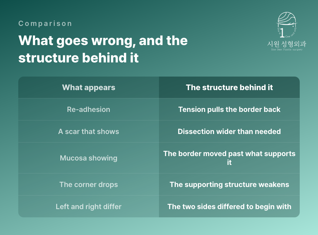
Re-adhesion is an opened area joining back together during recovery. It is the most commonly discussed of these, and the narrower the room left beyond the outer corner, the stronger the force pulling it back.
That is why what is done during surgery matters. Approaches that use a pretarsal fasciomuscular flap or a graft to deal with adhesion in revision surgery are reported separately in the plastic surgery literature — clearing tissue that wants to join is a technique in its own right.
Following the course through recovery affects the result too. The force to rejoin is strongest in the first weeks, while the tissue is still soft, and how the shape settles then determines whether anything further is needed. Re-adhesion is not rare, so the process does not end with surgery; recovery is part of it.
What divides an eye that scars from one that does not?
A scar is governed more by the area dissected than by the length of the incision. A craniofacial surgery report covering 421 epicanthoplasties notes that dissecting more widely than necessary can leave a hypertrophic scar or a web-shaped deformity.
So the same technique gives different results. An eye with thin skin and low tension settles its border with a narrow dissection, while an eye with thick skin and a strong pull keeps load on the suture line even when the same extent is opened.
Why does mucosa come into view?
The red mucosa lining the inside of the eye normally sits hidden behind the lid. When a corner opening moves the border past the line the lid can support, the surface that should stay hidden comes forward into view.
Widening horizontally and widening vertically act along different axes. A report on canthoplasty in a plastic surgery journal likewise separates double-eyelid surgery, which changes the vertical dimension, from inner and outer corner openings, which change the horizontal. Widening only the horizontal without establishing which axis is lacking tends toward mucosa showing.
Once exposed, mucosa does not cover itself again with time. The border has to be returned to where the lid originally covered it, and the surrounding tissue repositioned in the process. That is how much weight the first decision carries about how far to move the border.
Can the corner drop, or turn outward?
Where the tendon holding the outer corner and the tissue around it weaken, the lower lid can roll outward. An ophthalmology report on a lower lid pulled down by scarring describes releasing the adhesion together with lid fixation and a skin graft. That is why reversing it becomes a larger operation than the first one.
Where the structure has already sunk and mucosa is showing, correcting only the inner side does not restore the balance. Rebuilding a collapsed outer corner is needed alongside.
Why do the two sides differ?
Two eyes are never identical to begin with. The thickness of the lid, the height of the outer corner and the distance to the outer bone all differ slightly from side to side. Applying the same range to both is exactly what makes the difference show.
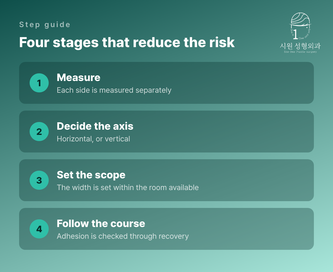
So the design is made separately for each side. How far to open on each is decided after measuring that side’s structure, and skipping that step is not infrequently what leads to a revision later.
Where the two sides have already settled differently, correcting only one is not the answer either. Which is possible with the tissue that remains — opening the less open side further, or returning the more open side — has to be established first.
How is the risk gauged before surgery?
Risk is gauged before surgery, not confirmed after it. Where the measured values fall on the side with little room, the plan changes to a narrower range or to looking at another axis alongside. The more sparing the width at a first operation, the more options remain later.
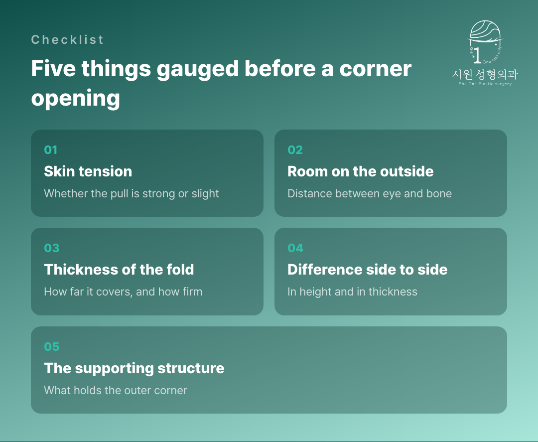
- Whether there is room between the eye and the outer bone — if narrow, the width of an outer-corner opening is limited.
- Whether the skin pulls strongly — if it does, load stays on the suture line for longer.
- Whether the epicanthal fold is thick and firm — if thick, the area dissected grows.
- How far the two outer corners differ in height and thickness — the design is made separately for each side.
- Whether what is needed now is horizontal or vertical — choosing the wrong axis leaves the result odd however far the range is extended.
Side effects of an outer-corner opening, set out by type
A three-part series on our official Naver blog.
Part I — a squared-off deformity at the outer corner ›
What to establish in consultation
Rather than comparing the names of techniques, it helps to ask which values of your own eye were measured, and what range those values pointed to.
To say that every side effect of a corner opening can be eliminated would not be true. But which risks grow in which eye can largely be gauged before surgery, and setting the range on the basis of that assessment is the best preparation available now.
Nobody can promise an outer-corner opening entirely free of complications, and we will not pretend otherwise.
What can be done is to establish beforehand which risks run higher in your particular eye, and to keep the scope inside what that eye can hold. Because the two sides are never identical, each is assessed on its own. We will show you which measurement pointed to which limit, so that the worry you walked in with has an answer you can check rather than one you have to take on trust.
Individual recovery times and results vary, and side effects can occur. Please consult our medical team before making any surgical decision.
WhatsApp ConsultationInternational patients welcome›
Official WebsiteProcedures and medical staff›
KakaoTalk ConsultationSend us photos and ask›
LINE ConsultationEasier if you are outside Korea›
DirectionsApgujeong Stn. · JJ Building 2F›
Consultations are held in English with the surgeon and our staff directly, not through an interpreter. Written enquiries in English are welcome.
2F, JJ Building, 19 Apgujeong-ro 28-gil, Gangnam-gu, Seoul, Korea
Mon–Fri 10:00 – 19:00 · Sat 10:00 – 16:00 · (KST, UTC+9)
Closed Sun & public holidays · +82-2-512-1889
